You are here
Shared Facilities
-
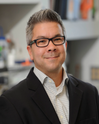
Mark J. Tomishima, Ph.D.
The SKI Stem Cell Research Facility
Sloan-Kettering Institute
Mark J. Tomishima, Ph.D.The SKI Stem Cell Research Facility provides the stem cell community with:
- Training in pluripotent stem cell culture, directed differentiation and transgenesis
- A collection of human embryonic and induced pluripotent stem cells (iPS)
- Customized iPSC production and characterization
- Shared equipment, including: flow cytometry, high content imaging, single cell isolation (C1), high content digital PCR, droplet digital PCR and conventional quantitative PCR.
- Screening small libraries of pathway activators and inhibitors
- Transitioning research-grade protocols to clinically-relevant GMP standard operating procedures (SOPs), with follow up manufacturing in the Cell Therapy and Cell Engineering Facility (Isabelle Rivière).
Website: http://stemcells.mskcc.org
E-mail: tomishim@mskcc.org -
Michael J. Fiske, M.S.
Upstate Stem Cell cGMP Facility
University of Rochester Medical Center
Michael J. Fiske, M.S.The newly constructed Upstate Stem Cell cGMP Facility (USCGF) is part of the URMC Stem Cell and Regenerative Medicine Institute and is designed as a multi-use cGMP manufacturing and testing facility with the goal of accelerating “first-in-man” early phase clinical studies. Capabilities include: development of clinical-scale manufacturing processes, development of analytical methods for product characterization and release, GMP manufacturing and Quality Control release testing of clinical-grade materials. The USCGF is available for contract manufacturing and testing services to both academic and private sector scientists.
Website: https://www.urmc.rochester.edu/upstate-stem-cell-facility.aspx
E-mail: mike_fiske@urmc.rochester.edu -
NeuraCell
Neural Stem Cell Institute
Steve LotzNeuraCell is a core facility that makes neural stem cell (NSC) research practical for those wishing to join the field. Our group has over 20 years of experience working with these cells and can now provide you with NSCs and everything you need to grow them. In addition to cells and media, we also provide a range of molecular tools including custom lentiviral shRNA and over-expression vectors. We also provide a consult based characterization service where we can assess how your products or reagents affect stem cell performance and behavior.
Website: http://www.neuracell.org
E-mail: stevelotz@neuralsci.org -

Gordana Vunjak Novakovic, Ph.D.
Stem Cell Functional Imaging Core
Columbia University
Nebo Mirkovic, Ph.D.The Columbia University Functional Imaging Core is a state-of-the-art facility that enables imaging studies for a variety of stem cell-related research areas. The equipment consists of a top-of-the-line Leica confocal / two-photon microscope, the CRi Maestro II small animal imaging system, an automated microplate reader, a live µCT imaging system, a high-throughput proteomics device, and an automated histology facility for tissue processing and embedding. The core is located at Columbia´s Morningside campus, and the space comprises equipment, imaging, cell culture and freezer rooms. Access is available to everyone for a per-usage fee, and booking is done via an on-line scheduling system.
Website: http://orion.bme.columbia.edu/gvnweb/nystem.htm
E-mail: nm2357@columbia.edu -

Paul Frenette, M.D.
Einstein Shared Facilities for Stem Cell Research
Albert Einstein College of Medicine
Paul Frenette, M.D.Einstein shared stem sell facilities provide services to promote pluripotent and somatic stem cell research, enhance epigenetic and genetic analyses in single stem cells, and facilitate in vivo stem cell transplantation.
Website: http://www.einstein.yu.edu/centers/stem-cell/core-facilities/
E-mail: paul.frenette@einstein.yu.edu -
John Schimenti, Ph.D.
Cornell Stem Cell Modeling and Phenotyping Core
Cornell University
John Schimenti, Ph.D.The Core provides state-of-the art capabilities for scientists to generate stem cells, modify genes in highly specific ways, create transgenic research animal models for basic and clinical research, analyze pathology of these animal models with high diagnostic and microscopic resolution, and study individual stem cells in live animals or in man-made environments.
The Core consists of three units:
- Stem Cell and Transgenics Core Facility
- Stem Cell Pathology Unit
- Stem Cell Optical Imaging Unit
The key feature of the Core is seamless via genome editing technologies from the experimental inception is tightly linked to characterization of these models at the gross pathological, histological, and cellular levels both in vivo and ex vivo. Investigators are encouraged to consult personnel of all three units at the stage of model design. Such an approach allows the most informed incorporation and efficient use of imaging and phenotypical modalities during the course of a research project.
Website: https://www.stemcell.cornell.edu/scp-mpcoreface.cfm
E-mail: jcs92@cornell.edu -
Richard Gronostajski, Ph.D.
Western New York Stem Cell Culture and Analysis Center
State University of New York (SUNY) at Buffalo
Richard Gronostajski, Ph.D.The WNYSTEM Stem Cell Center has 4 core facilities that provide:
- training in human embryonic (hES) and induced pluripotent stem (iPS) cell culture,
- generation of custom iPS cell lines,
- engraftment of stem cells in vivo in mouse models and analysis of their engraftment and
- next-generation sequencing of stem cells and their differentiated progeny using ChIP-seq, RNA-seq and other techniques for genetic and epigenetic analyses of cell function. Our goal is to promote and support stem cell research in western New York and beyond.
Website: http://wnystem.buffalo.edu/
E-mail: wnystem@buffalo.edu -

Marimar Lopez, Ph.D.
Rensselaer Center for Stem Cell Research
Rensselaer Polytechnic Institute
Marimar Lopez, Ph.D.The Rensselaer Center for Stem Cell Research (RSSCR) is a shared-use facility that provides state-of-the-art equipment, research collaboration and training for stem cell research. The RSSCR is open to investigators from Rensselaer Polytechnic Institute, (RPI), the Albany Capital Region and beyond. The Center offers hypoxic tissue culture and bioimaging equipment, several optical microscopy systems, epMotion robotics and sophisticated Thermo Arrayscan® and Olympus VivaView® imaging platforms. The Investigators using the RSSCR also are encouraged to utilize the science and engineering research cores adjacent to the stem cell facility, including micro-CT, MRI, wide-bore NMR, advanced microscopy (confocal, multiphoton, AFM, molecular capture/optical tweezers and TIRF), analytical biochemistry and FACS.
Website: http://stemcells.rpi.edu/
E-mail: lopezm4@rpi.edu -
Wadie F. Bahou, M.D.
Shared Facilities for the Stony Brook Stem Cell Center
Stony Brook University
Wadie F. Bahou, M.D.The Stony Brook Stem Cell Facility is located within the 250-acre Stony Brook Research and Development Park, uniquely developed for infrastructure support and device/product commercialization emanating from stem cell research and biomaterials. Specialized services for stem cell-related research include:
- Stem cell processing, derivation, distribution, and cryostorage of up to 200,000 vials;
- Viral vector development and distribution including high-titer lenti/retrovirus, adenovirus, and adeno-associated (AAV) virus;
- Proteomic, genomic, bioimaging, and phenotypic characterization of stem cells, and
- Training and educational workshops relevant to stem cell generation and targeted cell differentiation.
Discounted user fees ensure highly competitive pricing for all stem cell supplies, and specialized screening equipment for phenotypic characterization of stem cells. Specialized equipment includes:
- A Leica TCS SP8 X confocal microscopy with white light laser source that perfectly matches the wavelength of any fluorophore, and provides for simultaneous monitoring of eight excitation lines; and
- A Perkin Elmer High-Content Imaging System (Operetta) for multi-well image capture and monitoring, with remote user access seats for robust software analysis;
- Specially-retrofitted incubators for hypoxic growth and manipulation of stem cells.
Website: Stony Brook Stem Cell Center
E-mail: Wadie.Bahou@stonybrookmedicine.edu -

Shared Facilities and Resources for Stem Cell Research at The Rockefeller University and Weill Cornell Medical College
The Rockefeller University/Weill Cornell Medical College
Ali Brivanlou, Ph.D.At The Rockefeller University, Core Facility
- Followed the derivation of new hESC lines, RUES3 (Rockefeller University Embryonic Stem Cell Line 3), with the design of a genetically engineered human somatic fibroblast cell line that will be used as a platform for compounds screens. At Weill Cornell Medical College, Core Facility
- Progressed in pre-clinical experiments to set the stage for human trials, with advances made for clinical scale generation of hESC-derived endothelial cells and hematopoietic cells. Core Facility
- At The Rockefeller University Core progressed in chemical genetic screens to identify compounds able to direct differentiation towards neural lineages. Core Facility
- At Weill Cornell established a stem cell metabolite profiling facility to service the needs of investigators.
Website: http://rues.rockefeller.edu/
E-mail: brvnlou@rockefeller.edu -
Ihor Lemischka, Ph.D.
Human Embryonic Stem Cell (hESC) Core at Mount Sinai School of Medicine
Mount Sinai School of Medicine
Sunita D'Souza, Ph.D.The NYSTEM-funded, human embryonic stem cell (hESC)/induced pluripotent stem cell (iPSC) Shared Resource Facility (SRF) was established with the goal to promote hESC/iPSC research. The hESC SRF regularly conducts classes to teach iPSCs generation and hESC/iPSC differentiation into the lineage of choice. Scientists are also provided with tested stem cell reagents at vastly discounted pricing. To further the development of iPSC technology, the hESC/iPSC SRF is working to bring the latest innovations in the stem cell field to the scientific community to aid in the creation of a large number of transgene-free patient-specific iPSCs lines and differentiated lineages.
Website: http://icahn.mssm.edu/research/resources/shared-resource-facilities/human-embryo…
E-mail: sunita.d'souza@mssm.edu -
Lewis M. Brown, Ph.D.
Comparative Proteomics Center
Columbia University
Lewis M. Brown, Ph.D.At the Comparative Proteomics Center, we study proteins with differential occurrence in cells, tissues or affinity purified samples. NYSTEM funding with matching funds from Columbia allowed us to acquire a NanoAcquity liquid chromatograph and a Synapt QTOF mass spectrometer. This equipment allows us to use a label-free technique for mass spectrometry-based proteomics. The versatile, sensitive methodology allows flexible experimental design, and is ideally suited to stem cell biology, and has been used effectively in several stem cell studies that we have completed.
Website: http://www.columbia.edu/cu/biology/resources/proteomics/
E-mail: Lewis.Brown@Biology.Columbia.edu -
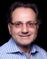
David Spector, Ph.D.
Confocal Microscope
Cold Spring Harbor Laboratory
David Spector, Ph.D.The confocal microscope purchased through NYSTEM funds allows CSHL scientists to visualize stem cells labeled with fluorescent dyes in order to estimate the number of stem cells, to observe the movement of stem cells and to observe differentiation of stem cells into specialized cells in living organisms.
Website: http://www.cshl.edu/Cancer-Center/Shared-Resources/Microscopy.html
E-mail: spector@cshl.edu
